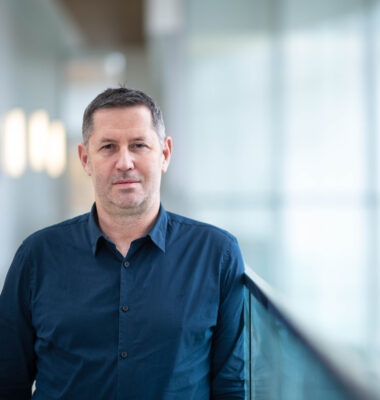
Alexander Rauscher
PhD
Associate Professor, Department of Pediatrics, Faculty of Medicine, UBC
Canada Research Chair in Developmental Neuroimaging
Full Member
Dr. Alexander Rauscher obtained his PhD in Physics from the TU Vienna, Austria, and after post-doctoral training at the Friedrich-Schiller-University Jena, Germany, he joined the UBC MRI Research Centre at the University of British Columbia in 2007. He became Assistant Professor at the Department of Radiology at UBC in 2010. In 2012, he received a CIHR New Investigator Award. In 2015 he joined the Department of Pediatrics at UBC as a Canada Research Chair in Developmental Neuroimaging.
Contact Info
2221 Wesbrook Mall
Vancouver, BC V6T 2B5
(Office located at G33 - Purdy Pavilion)
Research Information
How can we know whether a new treatment is able to repair a preterm baby’s brain? Or, how can we determine if a new drug protects the myelin sheath around nerve fibres from degradation due to multiple sclerosis (MS)? The answer is magnetic resonance imaging (MRI). Much of medical progress is propelled by new tools of observation running on MRI scanners. An MRI machine is like a smartphone that can’t do much without its ‘apps’ to fully explore the technology’s exquisite capabilities. The MRI scanner interacts with the human body via electromagnetic fields, “listens” how the body responds to these fields and, using lots of math and computational power, then translates the collected signal into meaningful images. Using this general principle, MRI scientists have learned how to map the nerve fibres in the brain, to make movies of the beating heart, or to detect changes in blood’s oxygen content as a marker for neural activity. This flexibility has made MRI an important technological platform for medical research and diagnostics.
My team of MRI scientists and I develop new MRI scans that measure brain tissue damage and how new treatments protect or even repair the brain. Such scans are urgently needed to measure the effectiveness of new treatments. I apply our new methods to MS and brain injury in newborns. With objective MRI markers we will be able to see treatment effects within weeks rather than months or years. MS is a disabling disease of the brain and spinal cord with no known cure. Its clinical course and symptoms are highly variable and unpredictable. 55,000 to 75,000 Canadians have MS. The ability to track tissue repair using MRI will be crucial for the evaluation of future drugs that are able to protect or repair the brain. Our sensitive MRI techniques will accelerate MS research and make drug trials cheaper.
In babies, on the other hand, we urgently need MRI scans that are able to accurately and objectively measure the effectiveness of new treatments. One in 100 infants is born before 34 weeks and the incidence is rising. While mortality has decreased, morbidity has not. Disability in preterm babies is almost exclusively due to damage to the very vulnerable developing brain. With objective MRI markers we will be able to see treatment effects within weeks rather than months or years. Our new imaging markers may lead to a dramatic acceleration of the search for effective interventions.
PublicationsKeywords
- fMRI
- MRI
- developmental neuroscience
- imaging
- multiple sclerosis (MS)
- Parkinson's disease
- concussion
- traumatic brain injury
